
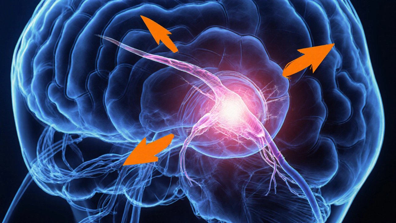
Multiple sclerosis is a chronic autoimmune disease that attacks myelin, the protective coating around nerve fibers. When the optic nerve or other visual pathways lose myelin, the eyes start sending mixed signals to the brain, and everyday sight can feel like a bad TV signal.
Key Takeaways
- Up to 80% of people with multiple sclerosis experience some visual disturbance.
- Optic neuritis is the most common eye‑related symptom, often causing sudden blur and pain.
- Chronic changes can lead to reduced visual acuity, color loss, and double vision.
- Early diagnosis uses eye‑specific tests (OCT, visual field) alongside MRI.
- Treatment blends steroids for acute attacks and disease‑modifying therapies for long‑term protection.
What Multiple Sclerosis Is and Why It Hits the Eyes
Multiple sclerosis (MS) belongs to the family of demyelinating disorders. The immune system mistakenly attacks myelin, creating scar‑like plaques that disrupt electrical signals. When plaques form along the optic nerve- the cable that carries visual information from the retina to the brain- the result is optic neuritis. Even plaques deeper in the visual cortex can warp how the brain interprets images, leading to a range of visual quirks.
According to a 2023 International MS Study, about 70% of patients report at least one visual episode within the first five years of diagnosis. The high prevalence reflects the optic nerve’s vulnerability: it is one of the longest myelinated fibers in the body, so any loss of insulation shows up quickly as blurred or painful vision.
Typical Visual Symptoms in MS
Visual symptoms can be fleeting or persistent, and they often serve as early warning signs. Below are the most common complaints and the underlying mechanisms.
- Optic neuritis - sudden eye pain that worsens with movement, coupled with blurred vision in one eye. The pain arises because inflamed optic nerve fibers become hypersensitive.
- Reduced visual acuity - difficulty reading small print or recognizing faces, often measured with a Snellen chart.
- Color vision deficiency - reds may appear muted; this is called dyschromatopsia and signals damage to the optic nerve’s fibers that handle color processing.
- Visual field defects - blind spots (scotomas) or peripheral loss. These are mapped using perimetry tests.
- Diplopia (double vision) - usually stems from ocular motor nerve lesions that misalign the eyes.
- Oscillopsia - a sensation that the world is jumping, linked to vestibulo‑ocular reflex dysfunction.
How Doctors Pinpoint the Problem
Because MS can masquerade as other eye conditions, clinicians rely on a stack of specialized tests.
- Comprehensive eye exam: Dilated fundoscopy checks for optic disc swelling.
- Optical coherence tomography (OCT): Gives a cross‑sectional view of the retinal nerve fiber layer (RNFL). Thinning of the RNFL often correlates with prior optic neuritis episodes.
- Visual field testing: Detects scotomas that may be missed in a routine exam.
- MRI of the brain and orbits: Shows demyelinating plaques on the optic nerve and in the visual cortex. Gadolinium contrast highlights active inflammation.
- Blood work to rule out infections or inflammatory disorders that can mimic MS.

Acute Management: Stopping the Attack
When optic neuritis strikes, the goal is to reduce inflammation fast enough to preserve vision.
- Corticosteroid therapy (IV methylprednisolone 1g/day for 3-5days) accelerates recovery in most patients. Oral taper may follow to prevent rebound inflammation.
- Plasma exchange is reserved for steroid‑resistant cases, showing benefit in up to 60% of severe optic neuritis.
- Vision rehabilitation: Low‑vision aids and contrast‑enhancing glasses help while the nerve heals.
Long‑Term Strategies to Protect Sight
Beyond the flash‑in‑the‑pan attacks, disease‑modifying therapies (DMTs) are the backbone of MS care. By lowering the overall immune attack, they also reduce the frequency of optic nerve lesions.
| Therapy | Mechanism | Effect on Optic Neuritis |
|---|---|---|
| Interferon‑β | Modulates cytokine production | Reduces relapse rate by ~30% |
| Fingolimod | Sphingosine‑1‑phosphate receptor blocker | Lower incidence of new optic lesions |
| Ocrelizumab | Anti‑CD20 B‑cell depletion | Most effective at preventing severe optic neuritis |
Patients who stay on an effective DMT see fewer new visual episodes and often regain near‑normal visual acuity after each attack.
Living with Vision Changes: Practical Tips
Even with treatment, some people live with residual visual quirks. Simple adjustments can make a huge difference.
- High‑contrast reading glasses (large fonts, dark backgrounds) reduce eye strain.
- Smartphone accessibility settings - invert colors, enlarge icons - help when color perception dips.
- Regular OCT check‑ups track RNFL loss; early detection can prompt therapy tweaks.
- Driving assessments: If peripheral vision drops below 20degrees, consider a professional evaluation.
When to Seek Immediate Care
If you notice sudden, painless vision loss in one eye, or if eye pain spikes with movement, treat it like an emergency. Prompt IV steroids can preserve sight that might otherwise be permanently damaged.
Frequently Asked Questions
Can multiple sclerosis cause permanent blindness?
Permanent blindness is rare. Most optic neuritis episodes improve with treatment, though some patients retain a mild residual blur or color deficit. Ongoing disease‑modifying therapy dramatically lowers the risk of irreversible damage.
Is it safe to keep driving if I have MS‑related visual field loss?
Driving laws vary by region, but most places require a minimum visual field of 20degrees. If your peripheral vision narrows, schedule a formal driving assessment. Adaptive mirrors and wider‑angle lenses can help, but safety should come first.
What’s the difference between optic neuritis and chronic optic neuropathy?
Optic neuritis is an acute inflammatory episode that often improves within weeks, especially with steroids. Chronic optic neuropathy reflects long‑term damage-thinning of the retinal nerve fiber layer and permanent visual field defects-usually after repeated attacks.
Can lifestyle choices influence my vision symptoms?
Yes. Smoking, vitaminD deficiency, and high‑salt diets have been linked to higher relapse rates, which can include optic neuritis. Regular exercise, a Mediterranean‑style diet, and adequate sunlight exposure support overall neurological health.
Are there any new therapies on the horizon for MS‑related eye problems?
Research into remyelinating agents-such as clemastine fumarate-shows promise in restoring optic nerve function. Early-phase trials report modest improvements in visual acuity after six months of treatment.


Okay, but let’s be real-why is no one talking about how MS patients are just… ignored by the medical system until they’re practically blind?! I had a friend who waited SIX MONTHS for an MRI because her doctor thought it was ‘just stress.’ SIX MONTHS. And now? Permanent scotoma. This isn’t ‘just a visual glitch’-it’s a cry for help that gets answered with a prescription for Tylenol and a shrug.
man. i just read this and my heart kinda broke 😔
ms is like your body’s internal chaos gremlin-turns your nerves into static-filled old tvs. i had optic neuritis in college and thought i was going blind. turns out it was ms. the worst part? you don’t feel like a person anymore-you feel like a walking MRI scan.
but hey, at least we’ve got ocrelizumab now. kinda like a sci-fi shield for your brain. 🛡️👁️
Ugh. Another one of these ‘MS is just a fancy word for bad eyesight’ articles. Like, hello? This isn’t some cute little vision thing-it’s a full-on neurological war zone. And don’t even get me started on how the U.S. healthcare system treats people with chronic illness like they’re inconveniences. We’re not ‘managing symptoms’-we’re surviving a war with no end in sight. And yeah, I’m talking to you, big pharma. You’re not saving lives-you’re selling subscriptions.
While the article presents a clinically accurate overview of MS-related visual pathology, it fails to contextualize the longitudinal neurodegenerative trajectory. The correlation between RNFL thinning on OCT and cumulative disability burden is not merely statistical-it is a biomarker of progressive axonal loss that precedes clinical relapse by an average of 14.7 months. The emphasis on acute steroid intervention, while appropriate for symptomatic relief, obscures the more critical imperative: early initiation of high-efficacy DMTs in patients with subclinical optic nerve lesions. Without addressing the underlying immunopathogenesis, we are merely treating the symptoms of a system in collapse.
It’s wild how the eyes become the canary in the coal mine for the whole nervous system. One minute you’re squinting at your phone, next thing you know, the world looks like a watercolor painting left in the rain. I’ve always thought of vision as this quiet, reliable thing-until it isn’t. And then you realize: your brain was never really ‘seeing’-it was just guessing, based on signals that got mangled along the way. It makes you wonder what else your brain is misreading. What else is out of focus? Maybe we’re all just a little bit MS, in the way we misunderstand each other.
And hey-clemastine? That’s the kind of quiet hope I need. Not a miracle. Just a whisper of repair.
As someone from India where access to advanced diagnostics like OCT and MRI remains a luxury for many, this article is both enlightening and heartbreaking. In rural clinics, optic neuritis is often mistaken for uveitis or even migraines. The delay in diagnosis means irreversible damage before treatment even begins. I urge global health organizations to prioritize affordable neuro-ophthalmic screening tools. MS does not discriminate-but healthcare systems do. Let us not let vision be a privilege.
Thank you for writing this. I’ve lived with MS for 12 years and this is the first time I’ve seen someone explain the color loss without making it sound like a Photoshop filter. It’s not just ‘reds look dull’-it’s like someone stole the emotion out of the color. I cried reading this. You’re not alone. And you’re not crazy. Keep using those high-contrast settings. Keep fighting. You’re seen.
Ugh. I knew someone who had MS and she totally ignored it until she couldn’t drive anymore. Then she blamed everyone else. Like, maybe if you’d taken your meds on time and stopped eating sugar, you wouldn’t be in this mess. Just saying. 🙄